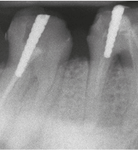
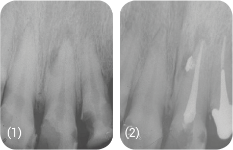
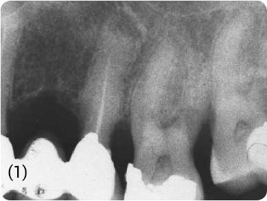
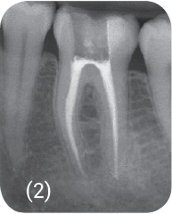
(1) Maxillary first molar: narrow canals and heavily mineralised dentine.
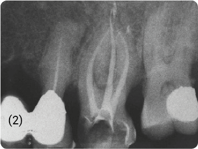
(2) Regular conicity obtained using CMA instruments.
Clinical case Pr. Roger Rebeiz
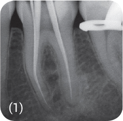
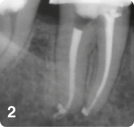
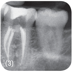
(1) Gutta points in place.
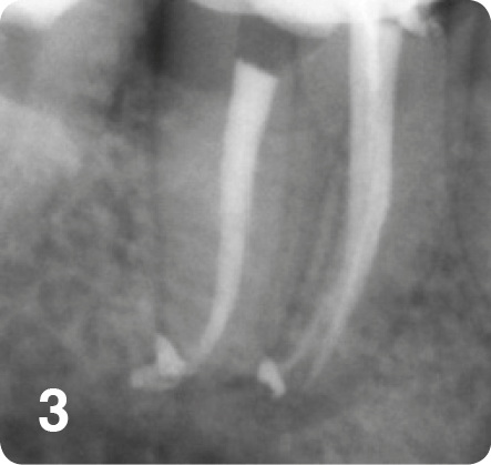
(2) (3) Root canal obturation seen from two different angles.
Clinical case Pr. Roger Rebeiz
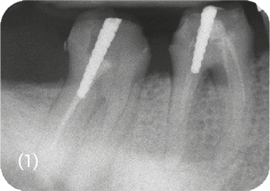
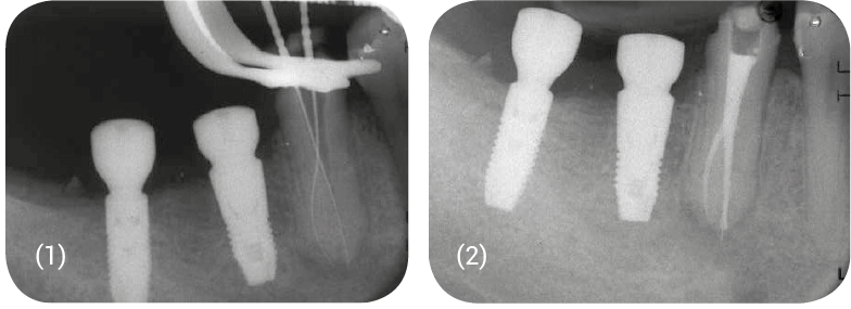
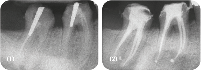
(1) Pre-op X-ray
(2) Post-op X-ray
Clinical case Pr. Roger Rebeiz
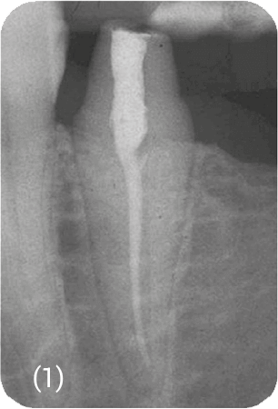
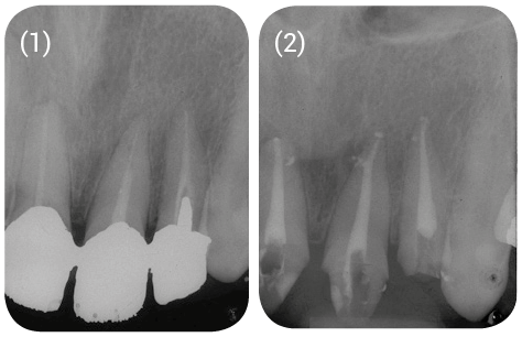
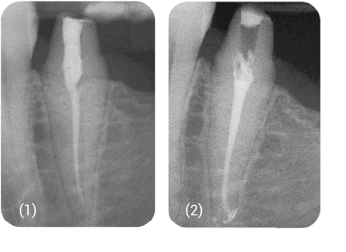
(1) Pre-op X-ray The access cavity must provide a clear view of the root canal entries and adequate access. • Cleaning out all traces of filling material. • Ultrasonic scaler is the technique of choice here. • Application of an appropriate solvent in the pulp chamber.
(2) Post-op X-ray
Clinical case Pr. Roger Rebeiz
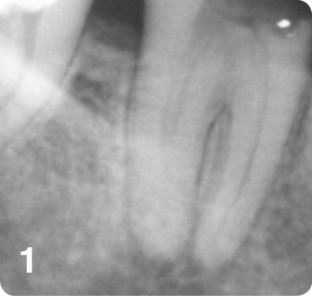
(1) Trajectories of the root canal prepared by PRESEQUENCE.